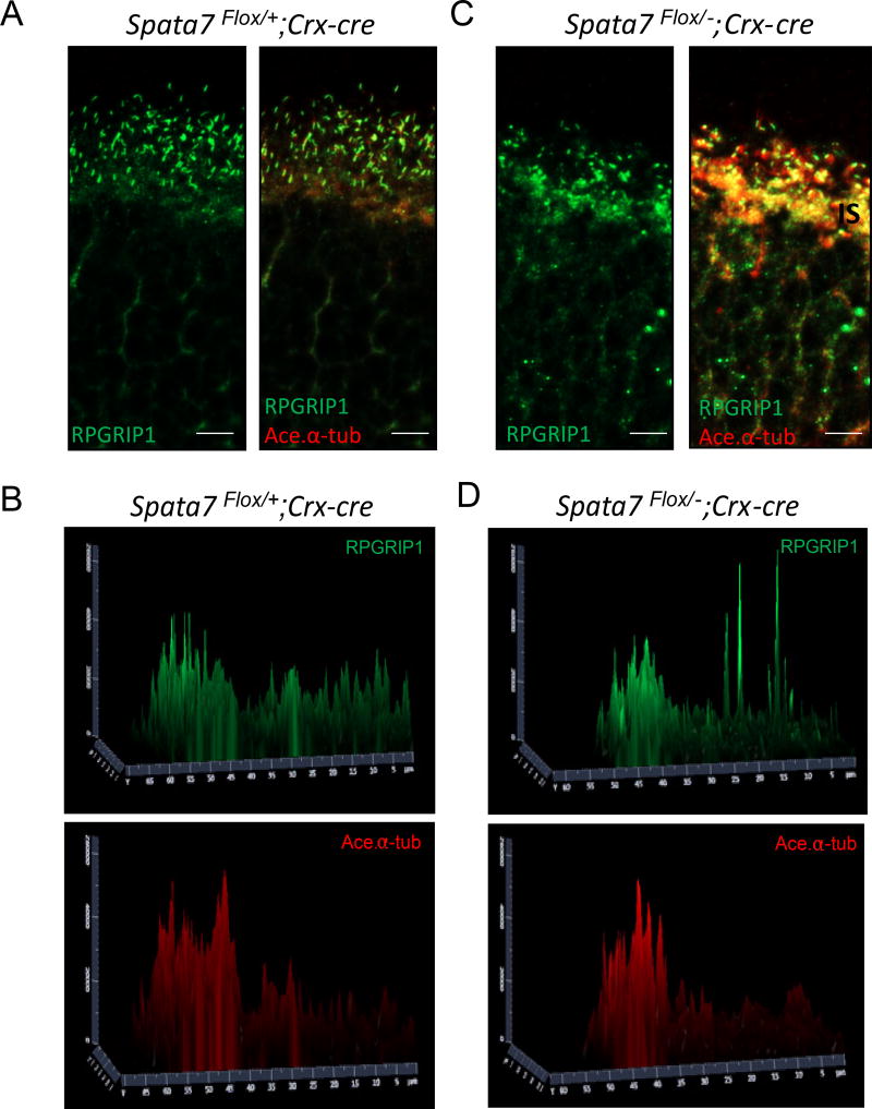
Figure 6. RPGRIP1 mislocalization in Spata7Flox/−; Crx-Cre retina.
(A) Immunostaining for RPGRIP1 was performed in Spata7Flox/+; Crx-Cre frozen retinal sections, where RPGRIP1 (green) co-localization was observed with acetylated-α tubulin (red) marker in the connecting cilium. (C) In Spata7Flox/−; Crx-Cre retinas, mislocalization of RPGRIP1 is evident, where it is detectable in the IS and ONL regions at P40. (B, D) Surface plot quantification of RPGRIP1 and Acetylated- α tubulin intensity in the OS, CC, IS, and ONL compartments. OS: outer segment; CC: connecting cilium; IS: inner segment; ONL: outer nuclear layer. Scale bar = 10 µm.

