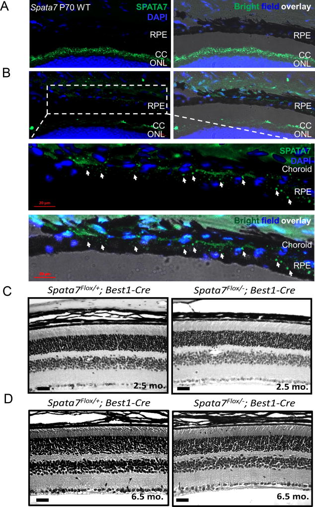
Figure 7. Spata7Flox/−; RPE-Cre conditional KO mice lack a retinal degeneration phenotype.
(A) SPATA7 expression (green) in the connecting cilium (left) and overlay of expression with bright field imaging of frozen mouse retinal sections (right). DAPI was used for nuclear counterstaining (blue). (B) Enhanced re-focusing of the SPATA7 signal in the RPE and choroid region at high magnification. Expression of SPATA7 (green) is indicated in the RPE by arrows. Scale bar= 20 µm. (C) Cross sections of 2.5-month-old RPE-specific Spata7 cKO retinas show no obvious photoreceptor degeneration and appear normal compared to Spata7Flox/+;Best1-Cre control retinal sections. (D) At 6.5 months, Spata7Flox/−; RPE-Cre and Spata7Flox/− ; RPE-Cre control mice consistently appear normal with no morphological defects present in retinas obtained from Spata7Flox/− ; RPE-Cre mice. RPE= retinal pigment epithelium, CC= connecting cilium, ONL= outer nuclear layer, OS= outer segment, INL= inner nuclear layer. Scale bar = 20 µm.

