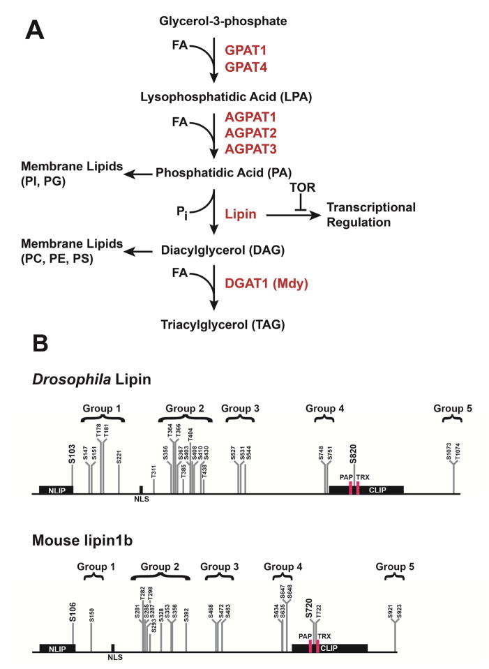
Fig. 1.
The lipogenic glycerol-3-phosphate pathway in Drosophila. (A) Overview of pathway and enzymes; FA, fatty acid; PI, phosphatidylinositol; PG, phosphatidylglycerol; PC, phosphatidylcholine; PE, phosphatidylethanolamine; PS, phosphatidylserine; (B) Comparison of mapped serine and threonine phosphorylation sites in Drosophila Lipin and mouse lipin 1b (Bodenmiller et al., 2008; Bridon et al., 2012; Harris et al., 2007). Sites are grouped in similar regions of the proteins. Two sites in the conserved NLIP and CLIP regions, which are highlighted by larger font, are homologous to one another. The locations of the catalytic motif, DIDGT (PAP), and of the transcriptional co-regulator motif, LGHIL (TRX), are indicated by red boxes; NLS, nuclear localization sequence.

