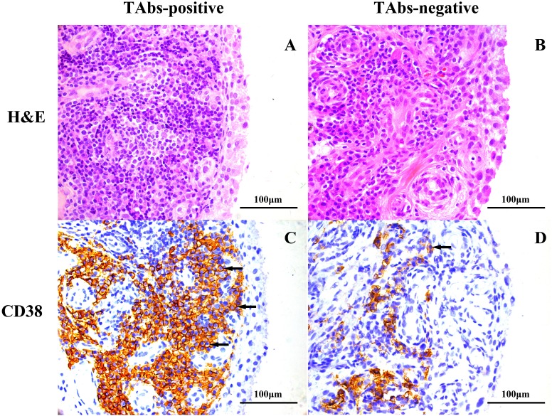
Figure 1. Representative images of H&E and immunohistochemical staining for CD38 in the synovium of RA patients based on TAb status.
H&E staining, high-grade synovitis (Krenn’s synovitis score = 6) in a TAbs-positive RA patient (A) and low-grade synovitis (Krenn’s synovitis score = 3.5) in a TAbs-negative RA patient (B); immunohistochemical staining, expression of CD38 in synovium of a TAbs-positive RA patient (C) and a TAbs-negative RA patient (D). Significantly more pronounced amounts of CD 38-positive plasma cells infiltrated the TAbs-positive synovium versus the TAbs-negative control. The black arrows point to CD38-positive cells located at the sublining area of the synovium. (A–D), original magnification, ×400.

