An official website of the United States government
Here's how you know
Official websites use .gov
A
.gov website belongs to an official
government organization in the United States.
Secure .gov websites use HTTPS
A lock (
) or https:// means you've safely
connected to the .gov website. Share sensitive
information only on official, secure websites.
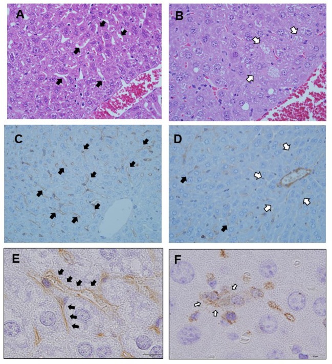
Figure 2. H&E and immunohistochemical analysis for the presence and localization of platelet aggregation and sinusoidal endothelial disorder after LPS administration. Liver sinusoids are lined with fenestrated endothelial cells and flanked by plates of hepatocytes in zone 3 immediately after LPS injection. Black arrows: Normal sinusoid (A). Sinusoidal dilatation and congestion occur in zone 3 at 12 h post-LPS injection. White arrows: Dilated sinusoid (B). Immunostaining with SE-1 immediately after LPS administration. Sinusoidal endothelial cells are stained (C). Immunostaining with SE- 1 at 12 h after LPS administration. Sinusoidal endothelial cells are not stained. Black arrows: Hepatic sinusoidal endothelial cells. White arrows: Lack of SE-1 staining in zone 3 (D). Immunostaining for CD62p immediately after LPS administration. Sinusoidal endothelial cells are stained. Black arrows: Hepatic sinusoidal endothelial cells (E). Immunostaining for CD62p at 24 h after LPS administration. Sinusoidal endothelial cells are not stained, and platelets are aggregated in the space of Disse outside the sinusoids. White arrows: Extravasated platelets (F).