. 2017 Nov 3;31(6):1103–1114. doi: 10.21873/invivo.11176

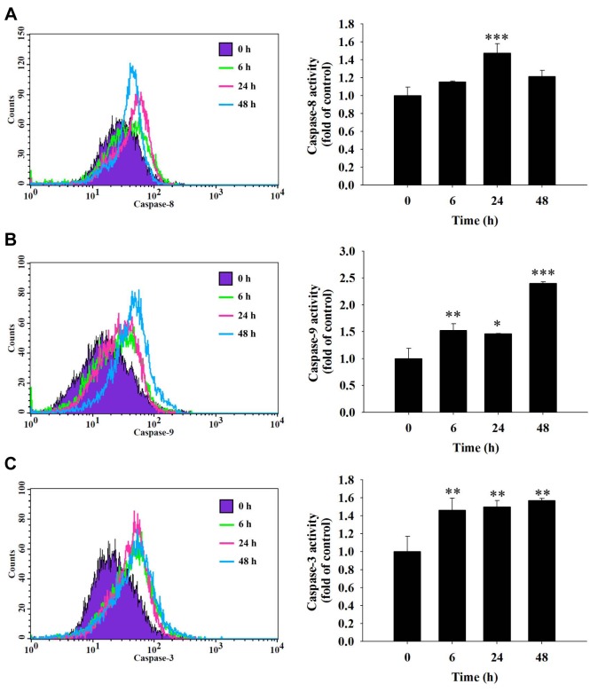
Figure 5. Fisetin induced the activity of caspase-3, caspase-8 and caspase-9 in HSC3 cells. HSC3 cells (5×10⁴ cells/ml) were plated in a 12-well plate and treated with 40 μM fisetin for 0, 6, 24 and 48 h then cells were harvested and re-suspended in 25 μl of 10 μM substrate solution containing CaspaLux8-L1D2 for caspase-8 (A), CaspaLux9-M1D2 for caspase-9 (B) or PhiPhiLux-G1D1 for caspase-3 activity determinations (C) before being incubated at 37˚C for 60 min. After incubation, all samples were washed with PBS and caspase activities were analyzed by flow cytometry as described previously. Significant differences at *p<0.05, **p<0.01, and ***p<0.001 between fisetin-treated groups and the control as analyzed by Dunnett test.

Figure 5