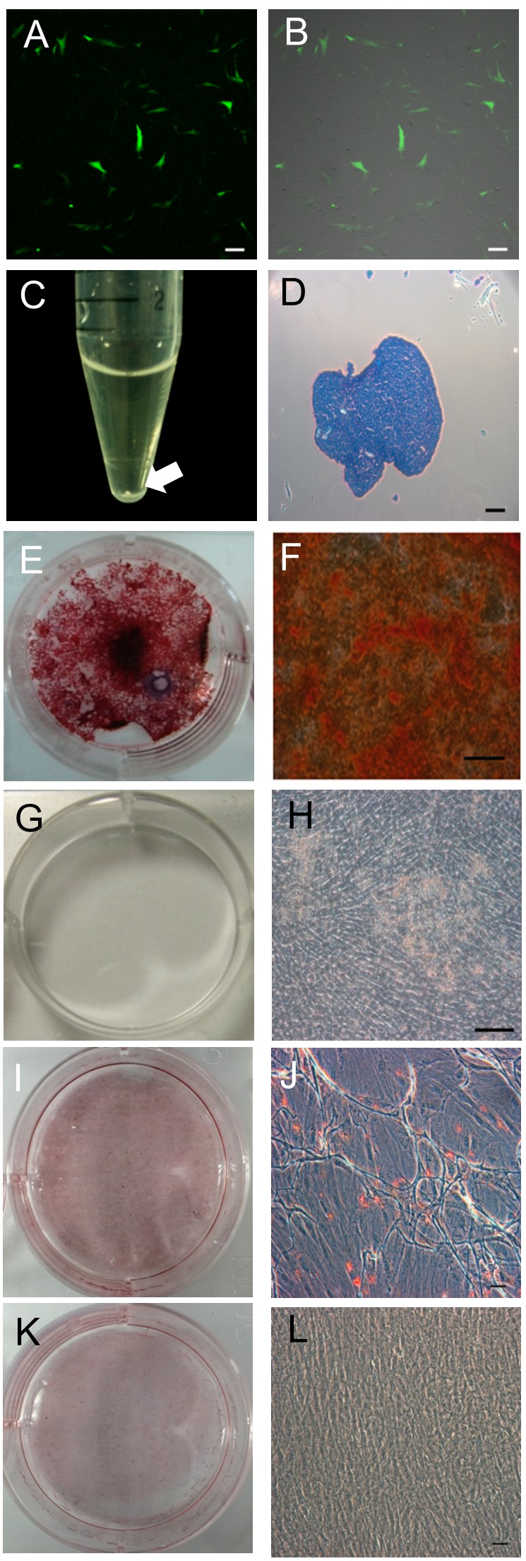
Figure 3. Characterization of canine umbilical cord blood-derived mesenchymal stem cells (cUCB-MSCs). A, B: cUCB-MSCs labelled with green fluorescent protein (GFP). A: Fluorescent microscopic image, B: merge with a light microscopic image, scale bar=100 μm. C, D: Chondrogenic induction: C: Pellet formation, aggregated to form a round shape. The white arrow indicates a pellet. D: Toluidine blue staining of chondrogenic pellets. The stained tissue showed a typical cartilaginous tissue phenotype. Scale bar=100 μm. E–H: Osteogenic induction: E, F: cUCB-MSCs grown in osteogenic induction medium. Differentiated cells stained strongly with Alizarin Red S, unlike control cells (G, H): scale bar=50 μm. I, J: Adipogenic induction. cUCB-MSCs grown in adipogenic induction medium. Differentiated cells stained strongly with Oil Red O, unlike control cells (K, L): scale bar=50 μm. I-L: cUCB-MSCs grown in maintenance medium: scale bar=50 μm.

