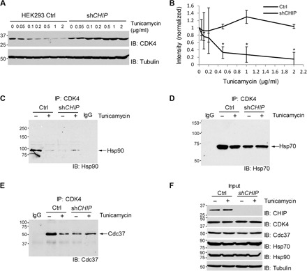
Fig. 10. ER stress–induced degradation of CDK4 is mediated by CHIP.

(A) shCHIP abrogates down-regulation of CDK4 protein in HEK293 cells after treatment with tunicamycin. Cells were incubated for 72 hours with tunicamycin at the indicated concentrations and harvested for immunoblotting. (B) Quantification of CDK4 protein levels after normalization by the levels of tubulin. Data are means ± SEM from three biological replicates, and the asterisks indicate statistical significance (P < 0.05). (C) ER stress decreases association of CDK4 with Hsp90 in HEK293 control cells but not in shCHIP cells. (D) ER stress does not significantly affect CDK4 association with Hsp70. (E) ER stress decreases association of CDK4 with Cdc37 in HEK293 control cells but not in shCHIP cells. (C to E) Cells were incubated with tunicamycin (1 μg/ml) for 24 hours and then 10 μM MG132 was added to the medium, followed by incubation for an additional 1.5 hours. Cell lysates were immunoprecipitated (IP) with CDK4 antibody, followed by immunoblotting for the indicated proteins. IgG, negative control for IP with normal IgG. (F) The levels of the indicated proteins determined by direct immunoblotting using the cell lysates before IP.
