Figure 2.
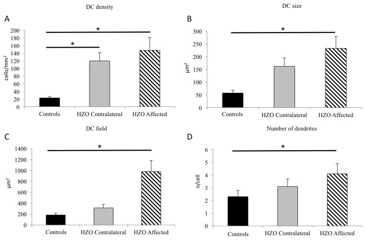
Dendritiform immune cell density (DCs) in herpes zoster ophthalmicus. Affected and contralateral eyes reveal statistical significant increase of DC density (A). DCs in the affected eye showed an increase in size (B), as well as DC field (C) and in number of dendrites (D). (* statistical significant adjusted p-value< 0.05)

