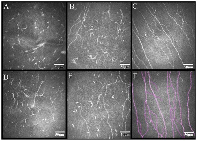
Figure 3.
Representative in vivo confocal microscopy images of the subbasal corneal nerve plexus in eyes with herpes zoster ophthalmicus and controls. Diminishment of nerve fibers is revealed in both affected eyes (A and D) and contralateral clinically unaffected eyes (B and E) of herpes zoster patients in comparison to normal controls (C). Example of nerve tracings performed by NeuronJ/ImageJ is shown (F).

