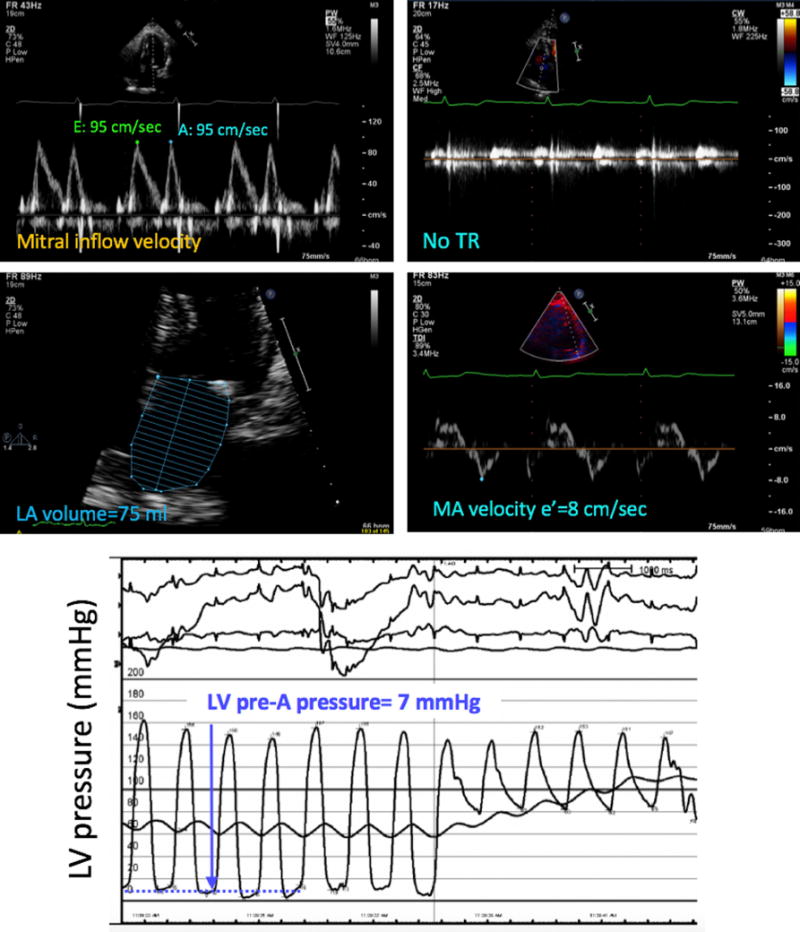
Figure 5.

Example of a patient in whom the noninvasive estimate of LV filling pressure (top four panels) did not match the invasive pressure measurement (bottom), which was normal. In contrast, measurements of individual echocardiographic parameters, as described in Figure 2, suggested elevated LV filling pressure using the 2016 ASE algorithm. MA, Mitral annular; Tr, tricuspid regurgitation.
