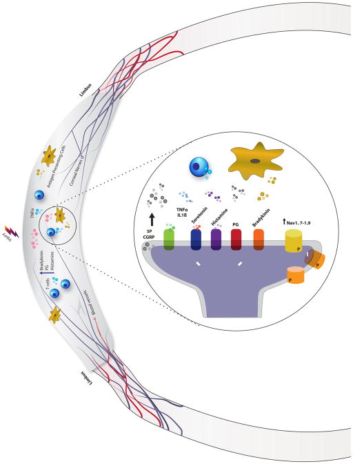
Fig. 1.
Mechanisms of peripheral sensitization. Injury to epithelial cells results in the release of numerous inflammatory mediators such as bradykinin, prostaglandins (PG), serotonin, and histamine, to name a few. Peripheral nerve terminals have receptors that recognize these inflammatory molecules (5HT for serotonin, H1 for histamine, EP for PG, B2/B1 for bradykinin). Their activation triggers the release of substance P (SP) and calcitonin gene-related peptide (CGRP). These mediators co-activate resident antigen presenting cells and recruit additional immune system cells to the site of injury. T cells and macrophages then secrete additional inflammatory cytokines (tumor necrosis factor alpha (TNFα) and Interleukin 1 (IL1)) that changes the function of peripheral nociceptors via protein kinase cascades. Mechanistically, this is largely prompted by the changes in ion channel function (through phosphorylation) and expression of new channels. These include specific ion channels such as voltage gated sodium channels (Nav1.7–1.9) and non-specific cation channels (transient receptor potential cation channel subfamily V member 1 (TRPV1).

