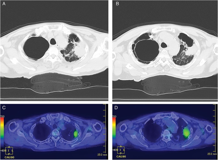
Figure 1.

(A–D) Computed tomography (CT) scan showing the nodule with marginal irregularity in the left upper lobe and the cavity with marginal infiltration in the right upper lobe (A, B). Positron emission tomography–CT scan revealing accumulated fluorodeoxyglucose concurrently with the nodule of the left upper lobe with pleural dissemination (C, D).
