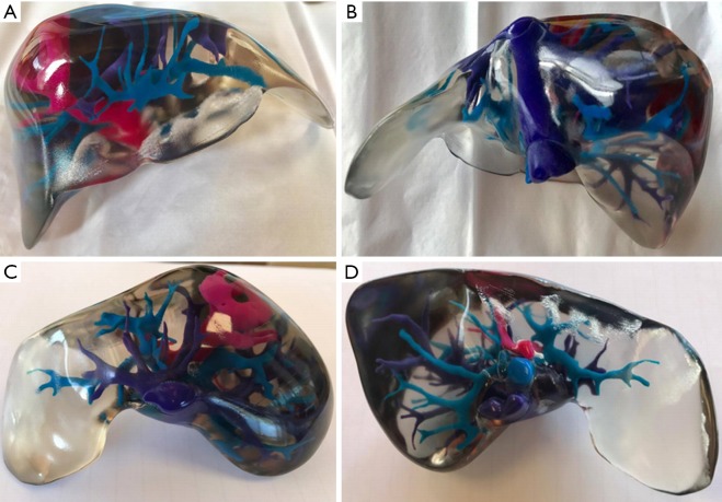
Figure 2.
Anterior (A), posterior (B), superior (C), and inferior (D) views of the 3D printed liver model generated from CT images, demonstrating the liver parenchyma (transparent), inferior vena cava and hepatic veins (purple), portal veins (blue), the tumour, and hepatic arterial supply (pink). 3D, three-dimensional; CT, computed tomography.

