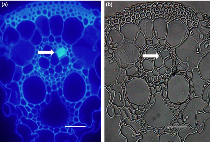
Figure 3.

Callose deposition in rice leaf sheath tissues obtained with fluorescence microscope at 40 × . White arrows show induced callose (with bright blue fluorescence) deposited on the sieve plates in the Si‐amended and BPH‐infested rice plants. (a) Obtained under ultraviolet. (b) Light microphotographs of a. Scale bar = 50 μm
