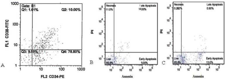
Figure 4.
Flow cytometric analysis of cord blood HSCs. (A) flow cytometric analysis of fresh CD34+ enriched cells. Specific staining was performed with anti-CD34 FITC and anti-CD38 PE antibodies. Flow cytometric analysis of apoptosis at day 14 in different culture condition of CD34+ with and without ADSCs feeder layer by PI and Annexin V staining. (B) cord blood CD34+ in co-culture with ADSCs, and (C) cord blood CD34+ without ADSCs feeder layer

