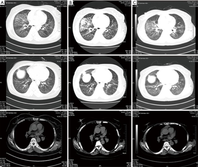
Figure 5.
Comparison of the chest CT. (A) 2014.6.27, after the treatment of six courses of INP. Nodules in the outer segment of right lung and multiple metastasis in lung hilars, mediastinal lymph nodes and thorax bone; multiple patchy shadows in the lungs; pericardial effusion absorbed; (B) 2014.11.10, 6 months after the treatment of six courses of INP. Progressed in the right lung lesions compared with the previous (2.1 cm × 1.9 cm), metastasis in lung hilars, mediastinal lymph nodes and thorax bone almost the same as before. Multiple patchy shadows in the lungs slightly increased; (C) 2015.6.12, after the treatment of S-1. Right lung cancer and right hilar and mediastinal lymph node metastasis; multiple interstitial and substance inflammation in the lungs decreased; multiple small nodules in the front left lung not clearly displayed, consider absorption. Little pericardial effusion absorbed. CT, computed tomography; INP, vinorelbine: 25 mg/m2, ifosfamide: 3,500 mg/m2 and cisplatin: 75 mg/m2.

