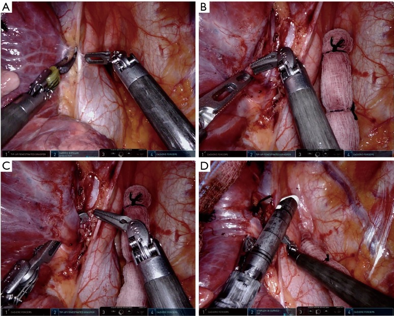
Figure 5.
Right upper lobe branch of superior pulmonary vein. (A) Tissue on the anterior surface of the vein is separated from the vein using the cadiere from port 4 and divided using the bipolar grasper from port 2; (B) blunt dissection using the cadiere from port 4 in the exit point of the stapler with the tip up fenestrated grasper from port 2 retracting the vein inferiorly; (C) passing of the tip up fenestrated grasper from port 2 with the cadiere from port 4; (D) vascular stapler from port 2 around the right upper lobe branch of the superior pulmonary vein.

