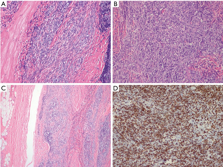
Figure 4.
Histopathology and immunohistochemistry. (A and B) Hematoxylin & eosin (H&E) stained sections of tumor, including regions showing (A) lymphocyte-poor spindle cell type A thymoma component and (B) lymphocyte-rich type B2 thymoma component (200× magnification); (C) H&E stained section of tumor capsule with focal area of intracapsular invasion (100× magnification); (D) TdT immunostaining highlighting immature T cells (200× magnification).

