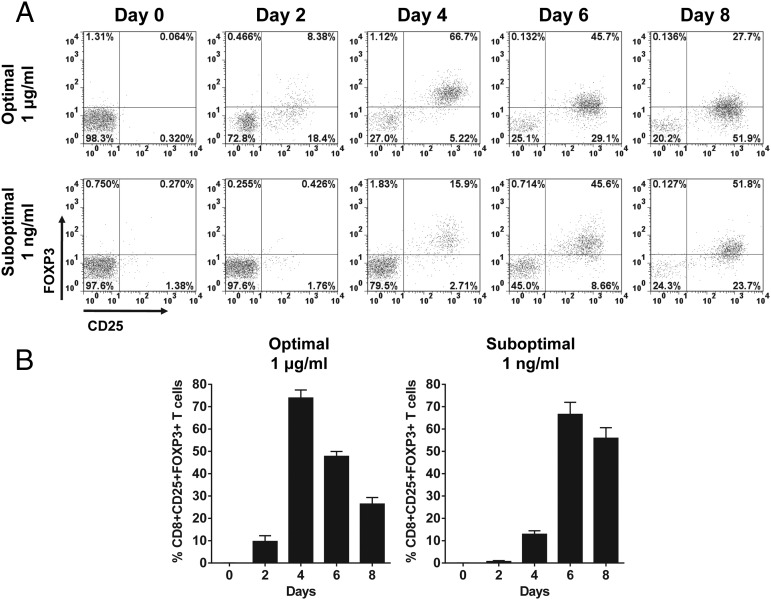
FIGURE 2.
Time-dependent expression kinetics of FOXP3 in CD8+CD25+ T cells induced by stimulation with SEC1. Human PBMCs were stimulated under optimal (1 μg/ml) and suboptimal (1 ng/ml) concentrations of SEC1 for up to 8 d. Expression of CD25 and FOXP3 in CD8 T cells was measured using flow cytometry. (A) Data shown are a single representative experiment gated on CD8+ T cells. (B) Percentage (mean ± SEM) of CD8+CD25+FOXP3+ T cells combined from nine independent experiments (three donors).

