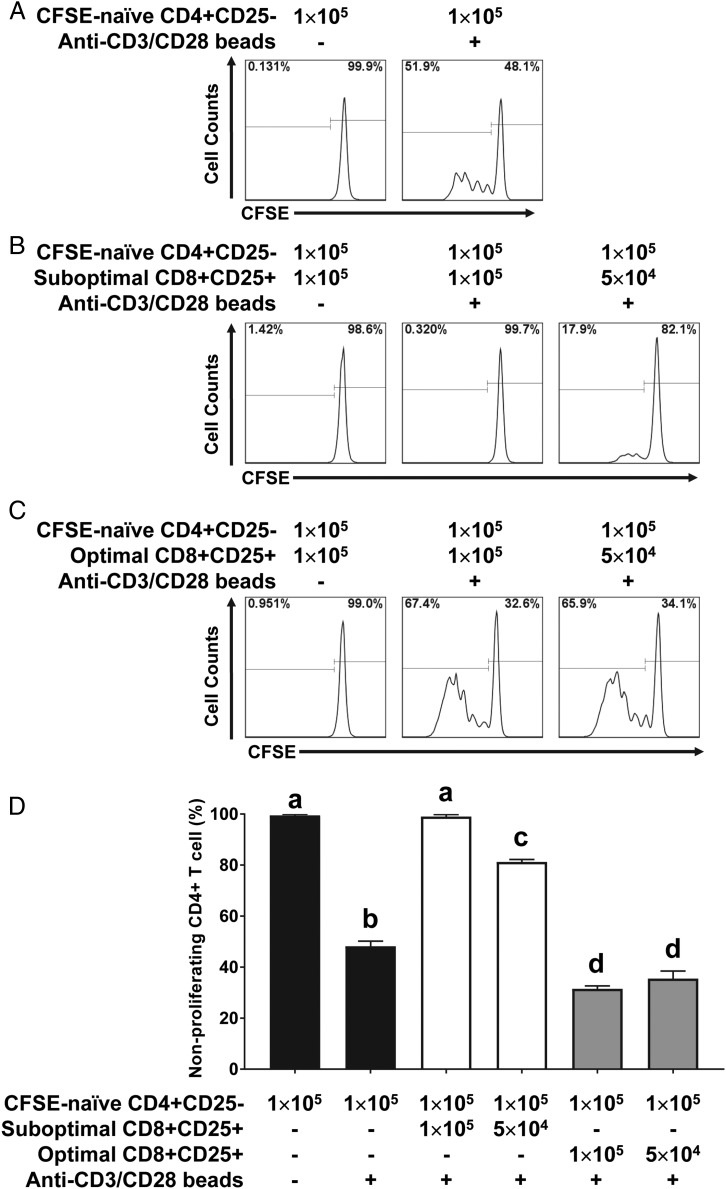
FIGURE 3.
Suppression of naive CD4+CD25− T cell proliferation by CD8+CD25+ T cells induced by suboptimal stimulation with SEC1. (A) Naive CD4+CD25− T cells were purified, labeled with CFSE, and stimulated with anti-CD3/CD28 beads for 4 d. Proliferation of CD4+ T cells was analyzed by quantifying the dilution of CFSE signal measured by flow cytometry. (B) CD8+CD25+ T cells induced by suboptimal stimulation with SEC1 for 6 d were purified and cocultured with CFSE-labeled CD4+CD25− T cells at various ratios for 4 d in the absence or presence of anti-CD3/CD28 beads. (C) CD8+CD25+ T cells induced by optimal stimulation with SEC1 for 4 d were purified and cocultured with CFSE-labeled CD4+CD25− T cells for 4 d in the absence or presence of anti-CD3/CD28 beads. (D) Percentage (mean ± SEM) of nonproliferating CD4+ T cells combined from nine independent experiments (three donors). Different letters indicate significant differences in the mean percentage between treatments, as determined by ANOVA, followed by the Tukey HSD test (p < 0.001).

