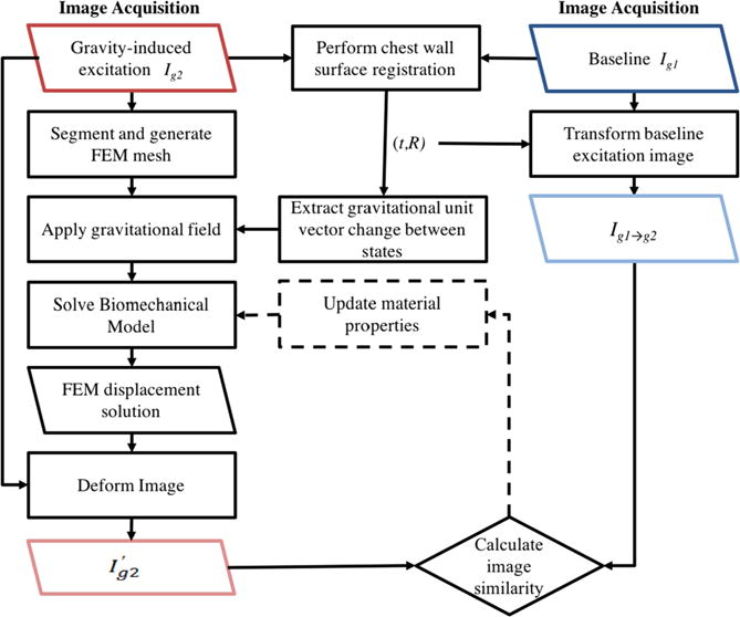
Figure 1.

The stiffness estimation framework begins with the acquisition of two gravity loaded image volumes (see figure 2). A rigid alignment between the two configurations is performed using only chest wall intensity information. The rigid registration procedure results in a translation, t, and rotation, R, that is used to transform Ig1 to be rigidly aligned with the chest wall in Ig2 (see figure 4). Also from R, a change in gravitational loading is quantified. A FEM mesh and biomechanical model is built from Ig2. A displacement field is generated from solving the biomechanical model and is used to deform Ig2. An image similarity metric is calculated between the model deformed image and the rigidly aligned baseline image (Ig1→g2). Stiffness properties are extracted when the calculated image similarity is optimized. An optimization procedure can be employed to iteratively update the material properties until the image similarity metric is minimized.
