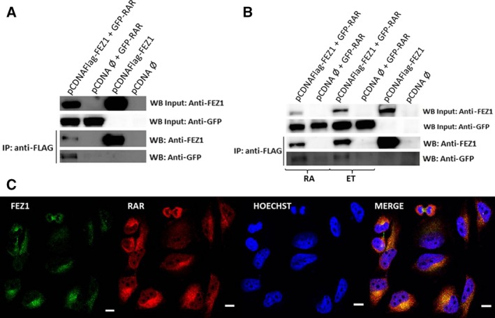
Figure 3.

FEZ1 and RAR interaction and localization in cells. (A) Overexpression of FEZ1 and RAR in COS cells and co‐immunoprecipitation using FLAG resin, followed by western blot. (B) Samples were treated with ethanol (ET; control) or all‐trans retinoic acid (RA, dissolved in ethanol) for 24 h and then co‐immunoprecipitated. (C) HeLa cells were analyzed by immunocytochemistry using anti‐FEZ1 (green) and anti‐RAR (red) antibodies and the nuclear dye Hoechst (blue). Colocalization occurred in the perinuclear region (yellow, merge image). White scale bar = 0.2 inches.
