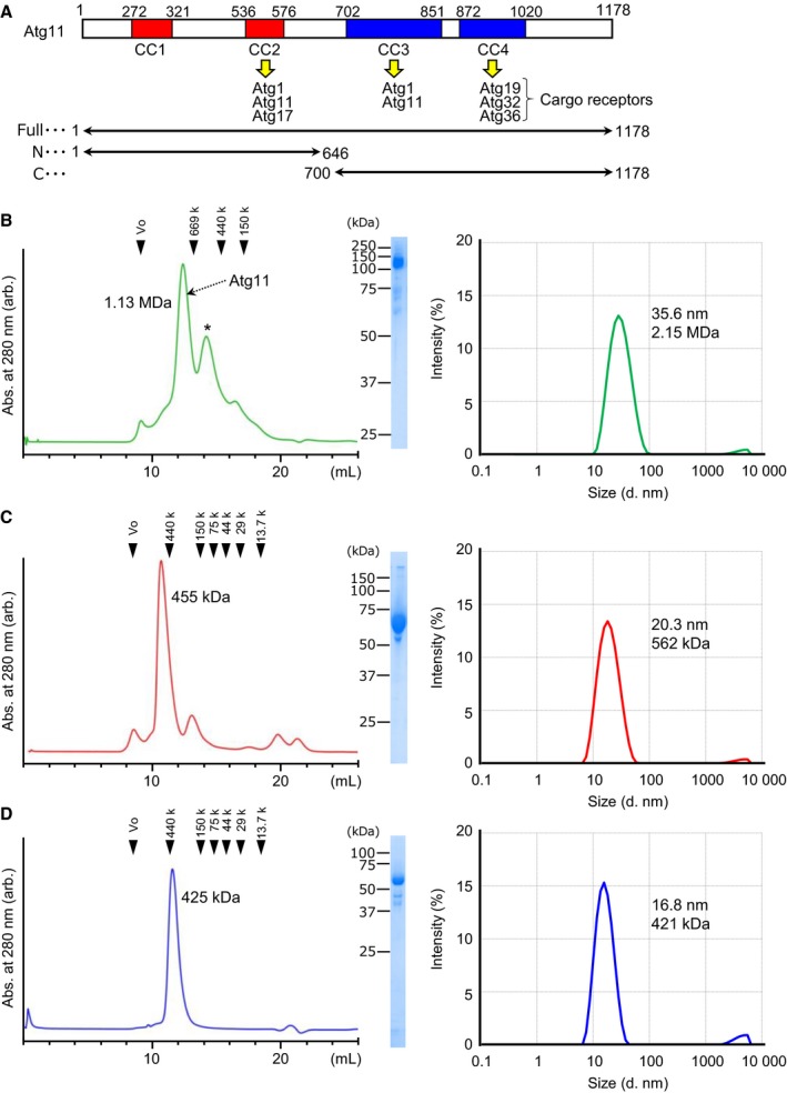
Figure 1.

Atg11 behaves as a monodispersed large particle in solution. (A) Summary of the known functional regions of Atg11. CC1–4 indicates predicted coiled‐coils, and each binding partner is listed. The constructs used in this study are shown below. (B–D) Elution profiles of the size exclusion chromatography are shown left, the SDS/PAGE pattern of the peak fraction is shown in the middle, and the size distribution of Atg11 measured by DLS is shown on the right. (B) Full‐length Atg11, (C) Atg11_N, and (D) Atg11_C. An asterisk indicates nonspecific or degraded proteins.
