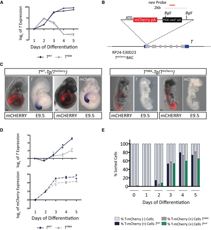
T expression in in vitro‐differentiated mesodermal cells from control T
WT and T
Y88A during the 5‐day differentiation protocol as measured by RT‐qPCR. n = 2 biological replicates.
Schematic illustrating the RP24‐530D23 T
mCherry reporter BAC. The orange line depicts the probe used to confirm genomic integration of the reporter BAC by Southern blot. Restriction enzyme sites used for Southern blotting are depicted.
Fluorescence images and WISH analysis of T
WT‐ and T
Y88A
‐Tg(T
mCherry) with T and mCherry probes at E9.5 reveals the co‐localization of T and mCherry expression from the T
mCherry reporter BAC. mCHERRY protein is more stable than the mCherry RNA and is localized to a broader domain. Scale bars: 200 μm.
T and mCherry expression in T
WT
‐ and T
Y88A
‐Tg(T
mCherry) cells differentiated into mesoderm in vitro during the 5‐day differentiation protocol, as measured by RT‐qPCR. The mean of n = 2 biological replicates is depicted.
Bar graph depicting the percentage of T‐mCHERRY(+) mesodermal cells and T‐mCHERRY(−) cells, as assayed by FACS, in T
WT
‐, T
Y88A
‐, and T
null
‐Tg(T
mCherry) cells during the 5‐day in vitro differentiation protocol.

