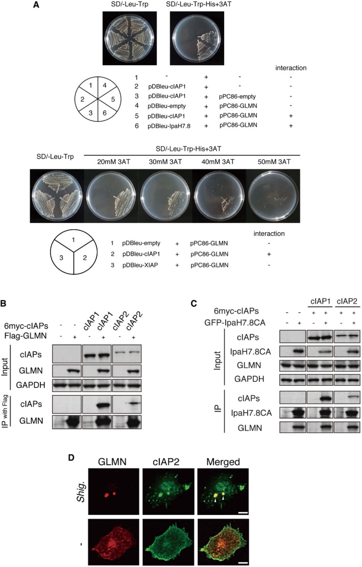
Figure 1. GLMN binds to cIAPs and forms a complex with Shigella IpaH7.8.

- Yeast two‐hybrid assay revealed direct binding between cIAP1 and GLMN. Saccharomyces cerevisiae strain MaV203 was transformed with the plasmid combinations indicated in the lower panel. The chart in the lower right summarizes the interaction results.
- Immunoprecipitation assay demonstrating the interaction between cIAPs and GLMN.
- cIAPs and GLMN co‐immunoprecipitated with Shigella IpaH7.8.
- Colocalization of endogenous GLMN (red) and cIAP2 (green) in BMDMs, visualized by confocal microscopy. WT BMDMs were infected with Shigella WT for 1 h and then subjected to immunohistochemical analysis using anti‐cIAP2 and anti‐GLMN antibodies. Scale bars, 10 μm. See also Fig EV1.
Source data are available online for this figure.
