-
A
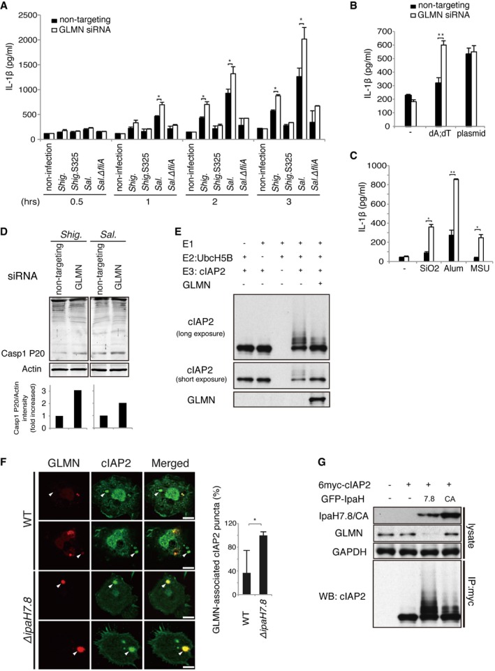
GLMN‐knockdown macrophages were infected with Shigella at an MOI of 5 or Salmonella at an MOI of 1, and IL‐1β levels were measured at the indicated time points. Non‐targeting siRNA was used as a control. n = 3, *P < 0.01.
-
B, C
GLMN‐knockdown cells were treated with stimulators of AIM2 (B) or NLRP3 (C) inflammasomes for 16 h, and then, levels of IL‐1β were measured. n = 3, *P < 0.01, **P < 0.005.
-
D
GLMN‐knockdown cells were infected with Shigella at an MOI of 5 or Salmonella at an MOI of 1, and then, the active mature form (P20) of caspase‐1 was detected by immunoblotting.
-
E
In vitro ubiquitination assay showing that GLMN inhibits self‐ubiquitination by cIAP2. Ubiquitination reactions were performed in the presence of purified ubiquitin, ATP, E1, E2 (UbcH5B), E3 (cIAP2), and GLMN.
-
F
Confocal imaging analysis of cIAP2 (green) and GLMN (red) at the late stage of Shigella infection. BMDMs from caspase‐1 KO mice were infected with Shigella WT or ΔipaH7.8 for 3 h, and then, the GLMN/cIAP2 complex was visualized by immunohistochemistry using anti‐cIAP2 and anti‐GLMN antibodies. White arrows indicate the localization of GLMN‐associated cIAP2 puncta. Scale bars, 10 μm. Graph indicates the ratios of GLMN‐associated cIAP2 puncta to total cIAP2 puncta in Shigella‐infected BMDMs, expressed as a percentage (%). n = 20, *P < 0.0001.
-
G
293T cells were transfected overnight with or without 6myc‐tagged cIAP2 and GFP‐tagged IpaH7.8 or IpaH7.8CA. 6myc‐tagged cIAP2 in cell lysates was immunoprecipitated with anti‐Myc antibody and subjected to immunoblot with anti‐cIAP2 antibody.
Data information: The error bars represent the SD of the measurements. Statistical analyses were performed using the Mann–Whitney
‐test. The results are representative of three similar independent experiments. See also Figs
.

