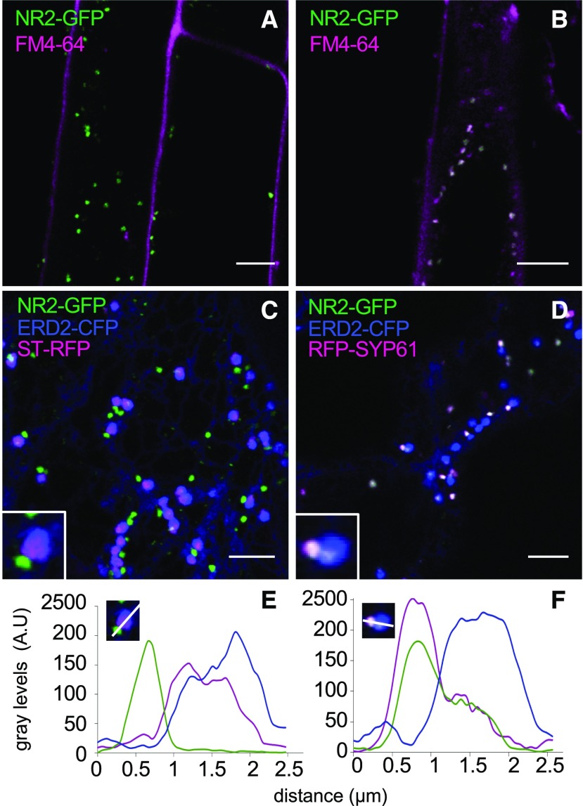
Figure 4.
NRAMP2 Is Localized to the TGN.
(A) and (B) Fluorescence pattern of the NRAMP2-GFP construct and kinetics of colocalization with the endocytic tracer FM4-64 ([A] 2 min; [B] 15 min) observed by confocal microscopy in roots of 5-d-old plants.
(C) and (D) Colocalization of NRAMP2-GFP with markers for cis-Golgi (ERD2-CFP), trans-Golgi (ST-RFP), and TGN (SYP61-GFP) transiently coexpressed in tobacco leaves. Inserts show a close-up image of the association of NRAMP2-GFP with the various Golgi subcompartments. Bars = 5 µm.
(E) and (F) Pixel intensity profiles of GFP (green), RFP (magenta), and CFP (blue) were measured along a line spanning the subcompartments magnified in the close-up insets shown in (C) and (D), respectively.

