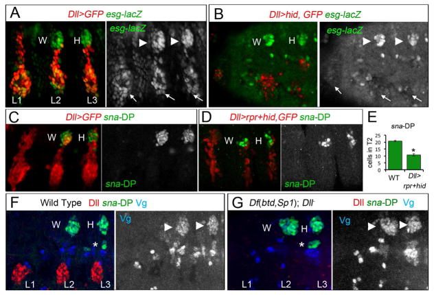
Figure 2. The DP is reduced when the VP is ablated.
(A) Thoracic view of esg-lacZ (green) and Dll>GFP (red) expression in wild type stage 14 embryos. The three leg and DP are labeled with arrows and arrowheads, respectively.
(B) Genetic ablation of the ventral primordia in Dll>hid embryos (red and arrows) reduced the size of the DP (arrowheads) and eliminates ventral expression of esg-lacZ, (arrows).
(C) Thoracic view of sna-DP (green) and Dll>GFP (red) in wild type stage 14 embryo.
(D) Ablation of the ventral primordia in Dll>rpr+hid embryos (red and arrows) reduced the size of the DP (green).
(E) Quantification of the number in T2 of sna-DP (green bars) in wild type and Dll>rpr+hid enbryos. * p<0.05 with Student’s t-test.
(F) Wild type stage 14 embryo stained for sna-DP-lacZ (green), Vg (blue) and Dll (red).
(G) A stage 14 Df(btd,Sp1); Dll− embryo. DP size is unaffected. An asterisk (*) in F and G label a band of muscle cells that express sna-DP and Vg. See also Figure S2.

