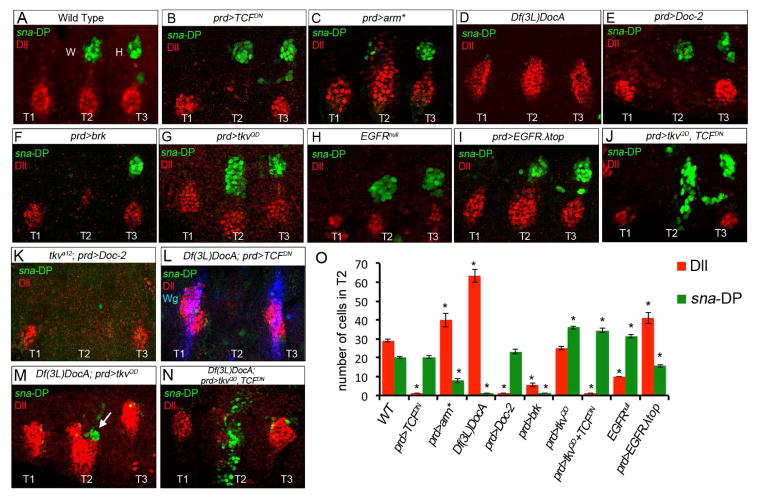
Figure 4. Distinct regulation of Dll and sna-DP.
Thoracic regions of stage 14 embryos stained for Dll (red) to mark the VP, sna-DP (green) to mark the DP, and Wg (L, blue).
(A) Wild type.
(B) In prd>TCFDN embryos the VP is absent and DP not affected in T2.
(C) In prd>arm* embryos VP size increases and DP size decreases in T2.
(D) In Df(3L)DocA embryos DP is absent and VP size is doubled.
(E) In prd>Doc-2 embyros, VP is absent and DP size is unchanged.
(F) In prd>brk embryos both the VP and DP are absent in T2.
(G) In prd>tkvQD embryos DP size increases, while VP size is unchanged in T2.
(H) In EGFRnull mutant embryos, VP size is reduced and DP size increases.
(I) In prd>EGFR. top VP size increases and DP size is reduced in T2.
(J) In prd>tkvQD, TCFDN embryos DP size increases and shifts them ventrally. VP are absent.
(K) A tkva12, prd>Doc-2 embryo. Without Dpp signaling, resupplying Doc-2 does not rescue DP formation.
(L) A Df(3L)DocA, prd>TCFDN embryo. Reducing Wg pathway activation in the absence of the Doc genes fails to rescue DP formation. Dll and wg expression are absent in T2.
(M) A Df(3L)DocA, prd>tkvQD embryo. Increasing Dpp pathway activity partially rescues DP formation (arrow).
(N) A Df(3L)DocA; prd>tkvQD, TCFDN embryo. Simultaneously activating the Dpp pathway and repressing the Wg pathway in the absence of the Doc genes further increases DP size (compare with M).
(O) Quantification of DP (green bars) and VP (red bars) size in the genetic backgrounds shown in (A–J). * p<0.05 with Student’s t-test indicates a significant difference from wild type T2.

