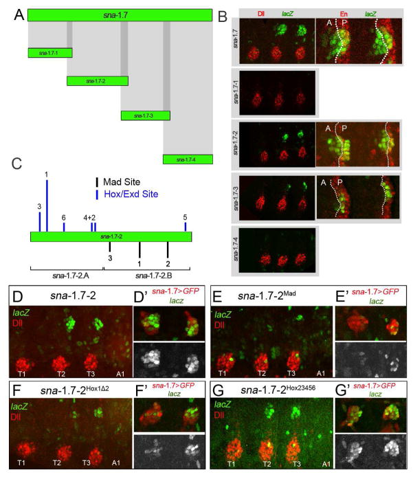
Figure 6. Molecular dissection of sna-DP.
(A) Division of sna-1.7 into 4 overlapping fragments.
(B) Thoracic segments of stage 14 embryos stained for Dll or En (red) and βgal (green). sna-1.7 reproduces the activity of sna-DP in both the A and P compartments. sna-1.7-2 is active in both compartments, while sna-1.7-3 is mostly restricted to the P compartment. sna-1.7-1 and sna-1.7-4 do not drive any expression in the DP.
(C) sna-1.7-2 has predicted binding sites for Hox/Exd (blue lines) and Mad (black lines). The height of the bar indicates the relative score (JASPAR). Subfragments sna-1.7-2.A and sna-1.7-2.B are indicated.
(D–G) Thoracic and first abdominal segments of stage 14 embryos stained for Dll (red) and βgal (green). The right panels show the DP region where the activity of sna-1.7>GFP (red) is compared to the activity of mutant sna-1.7-2 elements (green or white). (D) sna-1.7-2.
(E) sna-1.7-2Mad with the three predicted Mad sites mutated.
(F) sna-1.7-2Hox1Δ2 with Hox site 1 mutated. (G) sna-1.7-2Hox23456 with Hox sites 2–6 mutated. See also Figures S6 and S7.

