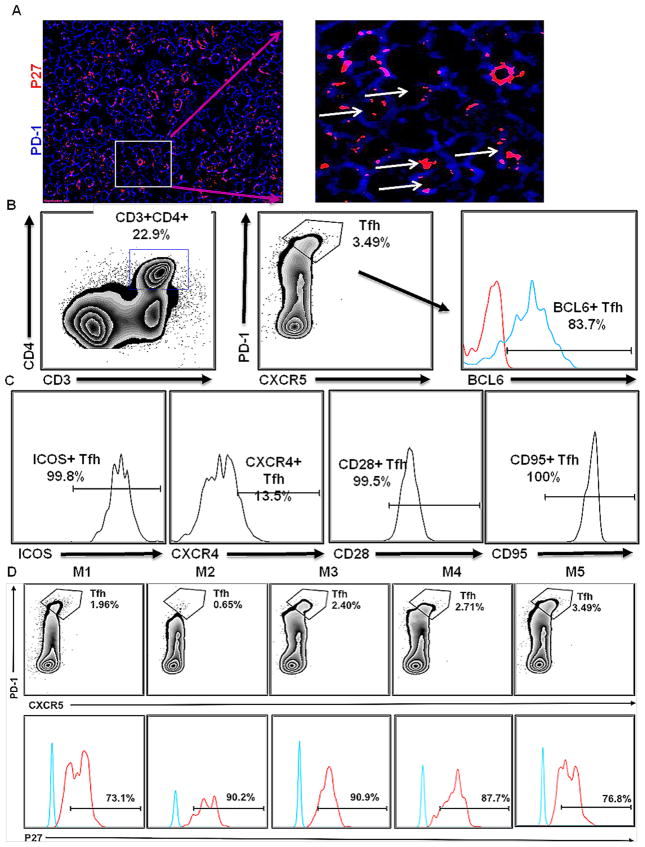
Fig. 6. Confirmation of SIV infection within TFHs of CLNs.
The use of confocal microscopy showing the presence of p27 (red) in PD-1hi TFH cells (blue) (A). The figure clearly shows intracellular antigen in TFH cells as shown by arrows in magnified image. Cells isolated from CLNs of chronically SIV-infected rhesus macaques (week 29) (n = 5) were characterized by FACS analysis. CD3+CD4+ cells were gated to select CXCR5+PD-1+ TFH cells (B). The phenotype of TFH cells was established using staining for BCL6, ICOS, CXCR4, CD95 and CD28 expression (C). The percentages of TFH cells in all rhesus macaque CLNs are shown in upper panel, while the percentages of TFH cells showing intracellular p27 are shown in lower panel (D). Blue and red line on the histograms corresponds to BCL-6 or p27 antibody and isotype control antibodies, respectively.

