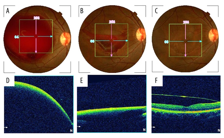
Figure 1.
Retina fundus photographs (A–C) and optical coherence tomography (D–F) of the right eye. An enlarged (8–10 disc diameters) preretinal hemorrhage located in the macula (A, D) at 31 weeks of gestation. Half of the hemorrhage was absorbed (B, E) 12 weeks after giving birth naturally. Complete reabsorption of the subhyaloid hemorrhage and a split premacular membrane were found in the macular region (C, F) nine month later.

