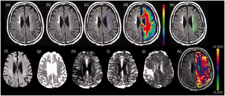
Figure 1.
Neuroimaging characteristics of diffuse microvascular dysfunction in a patient with acute ischemic stroke. Top row (panel a–e) demonstrates an example of the normal-appearing white matter region-of-interest in a patient with acute ischemic stroke and white matter hyperintensity volume of 2.36 cm3. (a) FLAIR image, (b) manual WMH outline (red), (c) dilated WMH outline (blue + red), (d) co-registered white matter probability mask, (e) contralesional normal appearing white matter mask (green) calculated as voxels with probability >95% of being white matter AND not in dilated mask (c). Bottom row (panel f–k) demonstrates this patient’s acute (f) DWI, (g) ADC, (h) CBF, (i) CBV, (j) MTT, and (k) K2 map shown overlaid on the FLAIR scan (for clarity only the contralateral hemisphere values are shown). All images are displayed in radiologic format.

