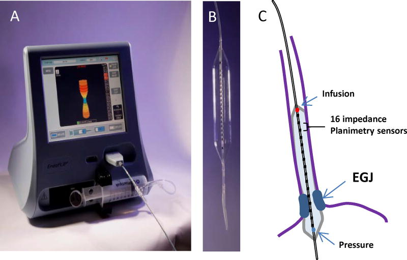
Figure 1. The functional lumen imaging probe (FLIP).
A) The EndoFLIP system (EF-100) with real-time 3-D imaging of the EGJ. The blue color on the screen represents the narrowest portion at the EGJ. B) An 10-cm balloon with 0.5-cm channel spacing housed within an 8-cm length FLIP segment (EF-325). C) Positioning of the 16 cm (EF 322) catheter with the distal portion through the EGJ and 10 recording segments in the body of the esophagus. The paired impedance planimetry rings (black) provide the measure of diameter and cross-sectional area. The pressure sensor (blue circle) is located in the distal aspect of the catheter and the infusion port (red circle) in the proximal aspect of the catheter within the balloon.

