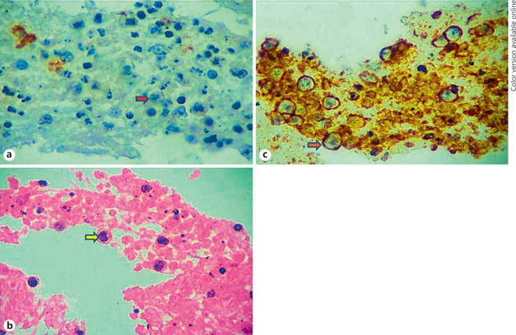
Fig. 1.
Cytopathology images of the patient. a, b Smears with hematoxylin-eosin and Giemsa stains show moderately cellular lymphoid cell population with abundant atypical lymphoid cells which are large, hyperchromatic with the chromatin less darker, prominent nucleoli, and scanty cytoplasm. Also seen are mature, small- and intermediate-sized lymphocytes accompanying these atypical lymphoid cells. The background shows fibrinous material. No granuloma is seen. c Immunohistochemistry shows atypical lymphoid cells with CD20 membrane positivity (orange arrow with blue outline).

