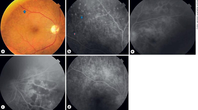
Fig. 2.
Color fundus and fluorescein angiogram findings in a patient with primary vitreoretinal lymphoma. a Color fundus image of the right eye showing multiple pinpoint deep, light yellow lesions spread across the retina. b Early-phase fluorescein angiogram image of the right eye showing multiple hyperfluorescent spots across the retina (blue arrow with blue outline) and multiple focal areas of capillary dropouts (red arrow with blue outline). c Large areas of capillary dropout seen nasal to the optic disc and extending inferonasally in the right eye. d Late-phase fluorescein angiogram image of the right eye showing diffuse granularity over the entire fundus. There is no flower petal leakage at the macula or disc leakage noted in the right eye. e Early-phase fluorescein angiogram image of the left eye showing diffuse granularity across the retina with no cystoid macular edema or disc leakage.

