Fig. 6.
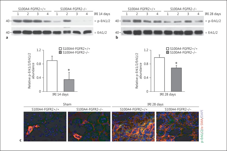
Erk1/2 phosphorylation was less in the kidneys from S100A4-FGFR2−/− mice after IRI. a, b Western blot analysis demonstrating the reduction in Erk1/2 phosphorylation in S100A4-FGFR2−/− kidneys at 14 days (a) and 28 days (b) after IRI compared to their control littermates. Numbers indicate individual animals. The gels were run under the same experimental conditions. The graphs show the semiquantitative results for p-Erk1/2 in the kidney lysates from S100A4-FGFR2−/− mice and control littermates at 14 and 28 days after IRI. * p < 0.05 versus control littermates, n = 4. c Representative immunohistochemical staining images showing the colocalization of α-SMA and p-Erk1/2 protein in fibrotic kidneys at 28 days after IRI. Asterisks indicate cells in the kidneys costaining positive for α-SMA and p-Erk1/2. α-SMA, alpha smooth muscle actin; Erk1/2, extracellular regulated protein kinase 1/2; FGFR, fibroblast growth factor receptor; IRI, ischemia/reperfusion injury.

