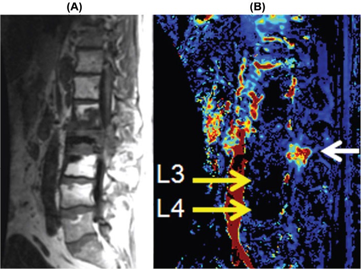
Figure 4.

Sagittal T1-weighted image (A) showing metastatic lesions at L1, L2, and L3 that were previously treated with radiation. The Vp color map (B) demonstrates recurrent disease involving the posterior elements at L2 (white arrow), which is indistinguishable from treated nonviable tumor at other locations. Note the postradiation changes of the marrow at L3 and L4 (high signal on the T1-weighted images) that are markedly hypoperfused (yellow arrows). The signal void (black region) in L2 body is polymethylmethacrylate cement from kyphoplasty for pathological collapse.
