
Figure 3.
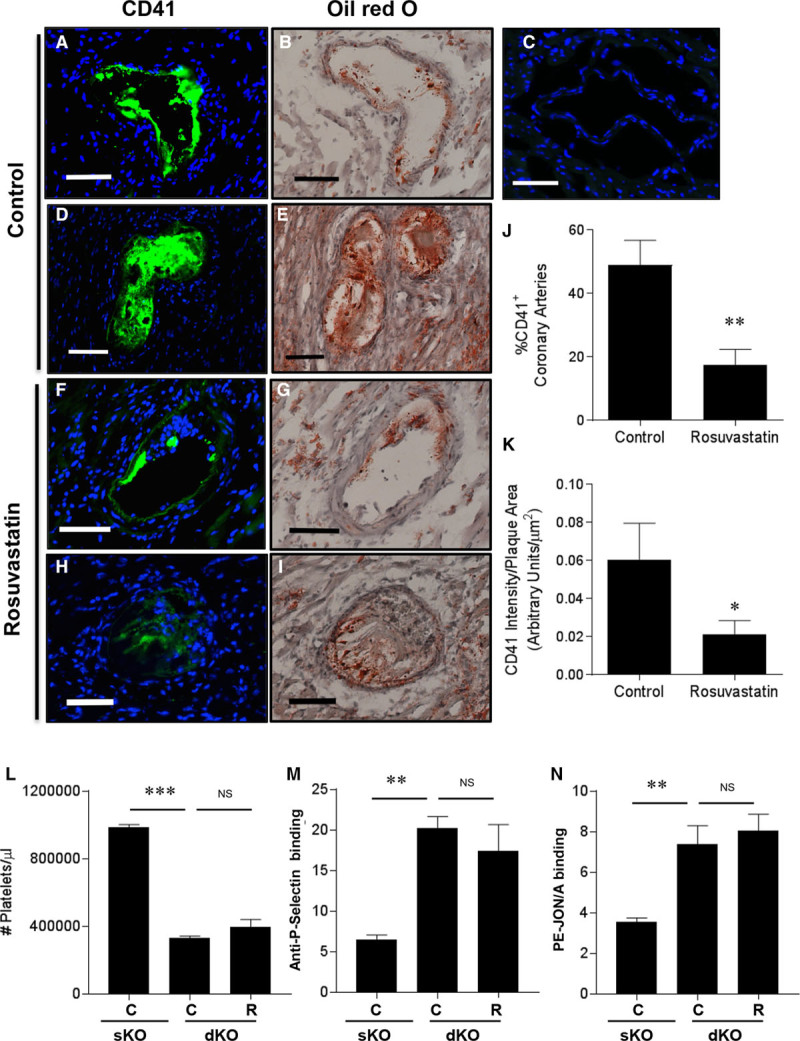
Rosuvastatin treatment reduced CD41 immunostaining in coronary arteries of SR-B1−/−/apoE−/− mice. Indirect immunofluorescence staining (green) for CD41, a marker of activated platelets, is shown in A, D, F, and H; (C) no primary antibody control (nuclear DNA stained blue with DAPI). Oil red O/hematoxylin-stained adjacent sections are shown in B, D, F, and H. A and B, Partially and (D and E) completely occluded coronary arteries from control saline-treated SR-B1−/−/apoE−/− mice. F and G, Partially and (H and I) completely occluded coronary arteries from rosuvastatin-treated SR-B1−/−/apoE−/− mice. Scale bar=50 µm. J, The percentage of CD41-positive atherosclerotic coronary arteries and (K) the intensity of CD41 staining (normalized to plaque area) in positive coronary arteries from control saline- (n=13) and rosuvastatin-treated (n=14) SR-B1−/−/apoE−/− mice. L, Circulating platelet counts, (M) anti-P-selectin antibody binding, or (N) PE-JON/A (phycoerythrin-labeled JON/A; anti-GPIIbIIIa) antibody binding to platelets from control apoE−/− (sKO) or control saline-treated or rosuvastatin-treated SR-B1−/−/apoE−/− (dKO) mice, detected by flow cytometry. Data in M and N are expressed as arbitrary fluorescence units. Data in J and K were analyzed by the Mann–Whitney rank sum test. Data in L through N were analyzed by 1-way ANOVA with the Kruskal–Wallis nonparametric test. *P=0.03, **P<0.01, *** P<0.002, and NS, not statistically significantly different vs control apoE−/− (sKO) or control saline-treated SR-B1−/−/apoE−/− (dKO) mice. apoE indicates apolipoprotein E; C, control saline-treated SR-B1−/−/apoE−/− (dKO) mice; dKO, double knockout; R, rosuvastatin-treated SR-B1−/−/apoE−/− (dKO) mice; sKO, single knockout; and SR-B1, scavenger receptor class B type 1.