
Figure 6.
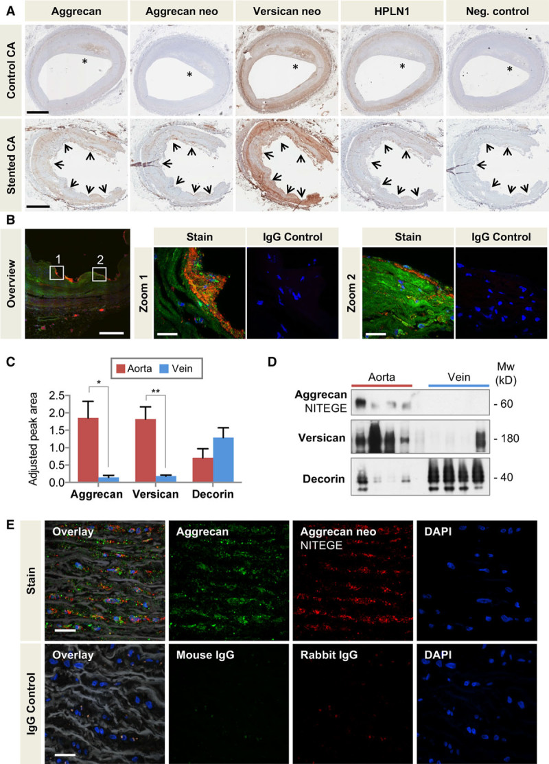
Aggrecan in the human vasculature. A, Localization of aggrecan, the aggrecan NITEGE, and versican DPEAAE neoepitopes (neo), and HPLN1 in stented and control human coronary arteries with the presence of atherosclerosis (*). Arrows mark the contacts of the stent struts with coronary artery. Scale bars=1 mm. B, Colocalization of aggrecan (Alexa 633, displayed in green) and aggrecan NITEGE neoepitope (Alexa 568, displayed in red) in human stented coronary arteries by immunofluorescence. Note the presence of aggrecan fragments at the contacts of the stent struts with the artery and in the subendothelial layer (zoomed-in areas). Overview image 20×, scale bar=500 µm; Zoomed-in areas 60×, scale bar=25 µm. C, Adjusted peak area for aggrecan, versican, and decorin in the human thoracic aorta and saphenous veins as determined by targeted proteomics. n=7 per group. *P<0.05, **P<0.01 (t test with unequal variance). D, Immunoblots for the aggrecan NITEGE neoepitope and versican in the human thoracic aorta and saphenous veins. The SLRP decorin served as a loading control. n=4 per group. E, Colocalization of aggrecan (Alexa 633, displayed in green) and aggrecan NITEGE neoepitope (Alexa 568, displayed in red) in human aorta visualized by immunofluorescence. Elastin fibers in white (autofluoresence with 488-nm laser excitation captured in the green emission channel). Control sections stained with isotope IgGs. Magnification 60×, scale bars=20 µm. CA indicates coronary artery; DAPI, 4′,6-diamidino-2-phenylindole; HPLN1, hyaluronan and proteoglycan link protein 1; IgG, immunoglobulin G; and SLRP, small leucine-rich proteoglycan.