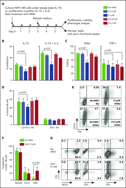
Figure 3.
Low-dose HMAs do not impair HSPC-NK cell viability, proliferation, and cytolytic functions. (A) Experimental design: Carboxyfluorescein diacetate succinimidyl ester–labeled HSPC-NK cells were cultured under proliferative (high-dose IL-15 and IL-2) or steady-state (low-dose IL-15) conditions in the presence of AZA or DAC refreshed daily at the indicated concentrations. Cell proliferation, viability, and absolute numbers, as well as functionality and phenotype, were analyzed by FCM after 6 days of treatment. (B) Percentages of proliferating HSPC-NK cells under steady-state (left panel) and proliferative (right panel) conditions. Combined data from 3 independent experiments (mean ± SEM) are shown. (C) Specific killing of K562 and THP-1 cells by HSPC-NK cells pretreated with HMAs. The same numbers of viable NK cells were plated in each experimental well after washout of the drug, and the killing of K562 and THP-1 cells was determined after overnight coculture using 1:1 E:T ratio. Data obtained with NK cells that were treated either under proliferative or steady-state conditions and performed with 6 different HSPC-NK cell donors are combined and depicted as mean ± SEM. (D-E) NK cell reactivity upon K562 stimulation and analyzed at the single-cell level by FCM. Combined data from 4 experiments using proliferative (n = 2) or steady-state (n = 2) conditions (D), and representative dot plots of HSPC-NK cells cultured under proliferative conditions and treated with AZA 1 µM, DAC 0.1 µM, or without HMAs (E) are shown. (F) Expression level of the maturation markers NKG2A, CD16, and killer immunoglobulin-like receptor-positive (KIR) cells on HSPC-NK cells following culture upon proliferative conditions in the presence of DAC 0.1 µM, or without HMAs. Mean ± standard deviation (SD) of 6 HSPC-NK cell donors is shown. (G) Representative dot plots of HSPC-NK cell IFN-γ production capacity with respect to KIR expression following DAC 0.1 µM or no HMA treatment under proliferative conditions. Statistical analyses were performed with 1-way (B-D) and 2-way ANOVA (E). NS, not stimulated.

