
Figure 5. Unlocking and triggering the primed Syt1-SNARE-Cpx-Syt1 complex.
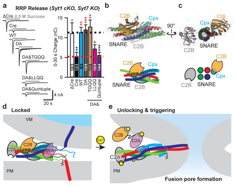
a, Recordings of IPSCs evoked by a 30-s application of 0.5 M hypertonic sucrose to induce exocytosis of RRP from cultured cortical neurons with Syt1 conditional KO and Syt7 constitutive KO. Cultures were infected with lentiviruses expressing ΔCre/Cre recombinase and WT Syt1 or Syt1 mutants. Sample traces (left) and summary graph of the IPSC charge transfers from RRP (right). Shown are means ± s.e.m; the number of neurons/independent cultures are indicated. Statistical significance was assessed by Student’s t test (*P < 0.05; **P < 0.01; ***P < 0.001) with respect to either the Cre (marked in red) or the Cre+Syt1 group (black). b and c, Orthogonal views (cartoon representation, upper; schema, lower) of the Syt1-SNARE-Cpx-Syt1 crystal structure with the membrane interacting elements of the primary Syt1/SNARE subcomplex located in a plane perpendicular to the page. d, Model of a primed Syt1-SNARE-Cpx-Syt1 (“Locked”) complex situated between the synaptic vesicle and plasma membranes. e, Upon unlocking and Ca2+-triggering, fusion occurs. For clarity, we omitted the transmembrane domains of the Syt1 molecules. Two or more such complexes are likely involved (e.g., Extended Data Fig. 9).