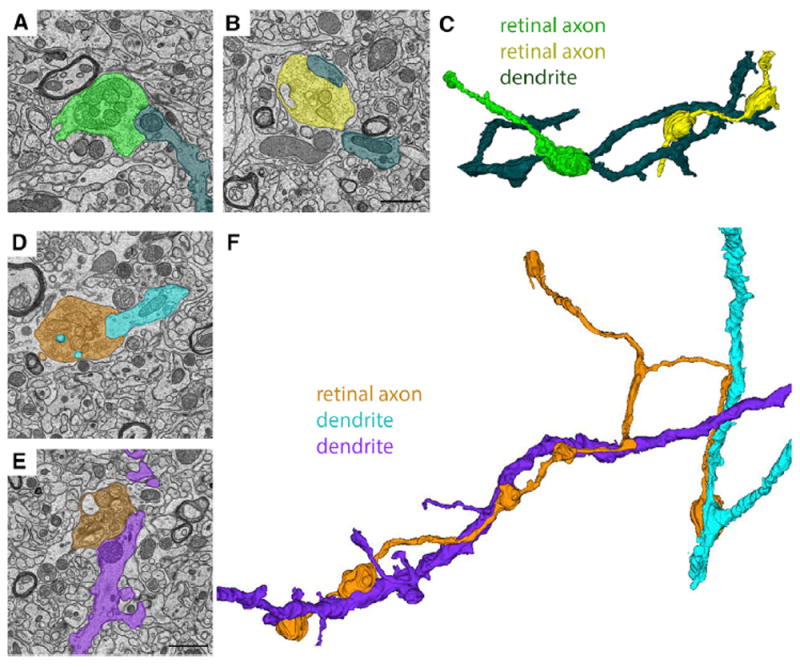
Figure 3. Ultrastructural Analysis and Reconstruction of Retinal Axons Contributing to “Simple Encapsulated” Retinogeniculate Synapses in dLGN.

(A and B) SBFSEM images of two retinal terminals synapsing onto the same relay cell dendrite in the “shell” region of dLGN.
(C) 3D reconstruction of the two RGC terminal boutons from (A) and (B) converging on a single relay cell dendrite.
(D and E) SBFSEM images of two retinal terminals from the same RGC axon making synaptic contact with two distinct relay cell dendrites in the “shell” region of dLGN.
(F) 3D reconstruction of the retinal axon and relay cell dendrites from (D) and (E). Scale bar in (B), 1.5 μm for (A) and (B), and in (E), 1.5 μm for (D) and (E).
