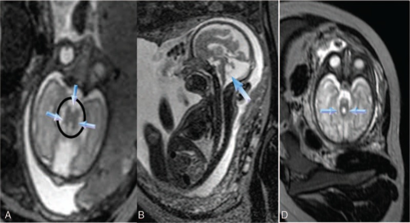
Figure 2.

(A) The molar tooth sign (circled) with a deeper interpeduncular fossa (down arrow); bilaterally thickened, elongated, and parallel superior cerebellar peduncles (SCPs) (other arrows), and a dilated cisterna magna are shown on axial T2-weighted imaging. (B) The enlarged fourth ventricle (arrow) remains on midsagittal T2-weighted imaging. (C) Two big curved SCPs (arrows) in another case. SCP = superior cerebellar peduncle.
