Figure 5.

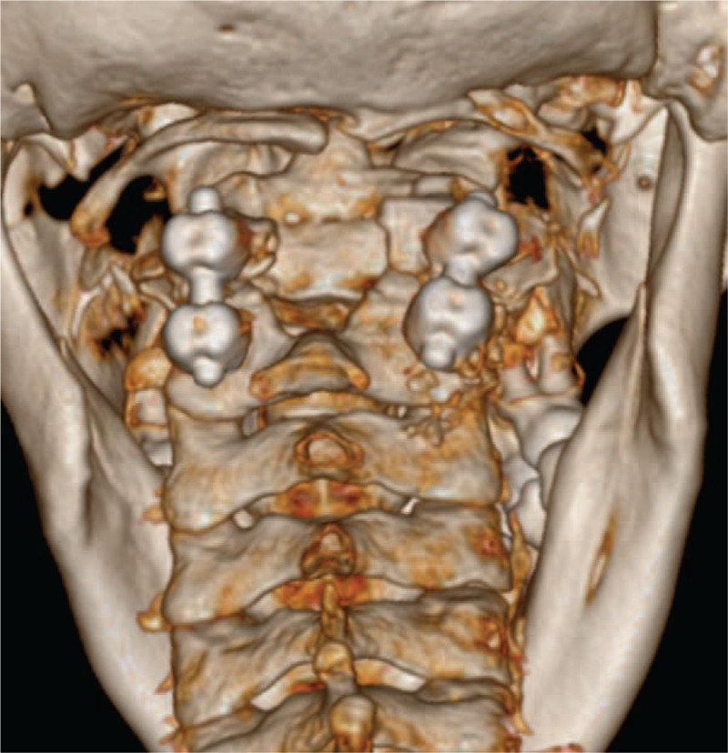
Postoperative three-dimensional CT image of the patients. A defect in the laminae of C2 and C3 was clearly shown after removal of the anomaly. CT = computed tomography.

Postoperative three-dimensional CT image of the patients. A defect in the laminae of C2 and C3 was clearly shown after removal of the anomaly. CT = computed tomography.