Figure 3.

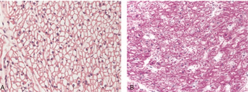
Photomicrography of the myocardium. A (H&E, 400×) and B (PAS, 400×), showing diffuse myocyte vacuolization. H&E = hematoxylin and eosin, PAS = periodic acid–schiff.

Photomicrography of the myocardium. A (H&E, 400×) and B (PAS, 400×), showing diffuse myocyte vacuolization. H&E = hematoxylin and eosin, PAS = periodic acid–schiff.