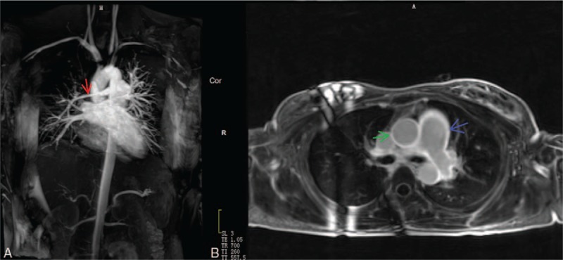
Figure 2.

A 22-year-old woman with the active Takayasu arteritis. Magnetic resonance angiography (MRA) (A) showed the stenotic right pulmonary artery (red arrow). Delayed contrast-enhanced magnetic resonance imaging (DCE-MRI) with three-dimensional inversion recovery Turbo fast low-angle shot (3D IR Turbo FLASH) (B) showed delay enhancement in aorta (green arrow) and pulmonary arteries (blue arrow).
