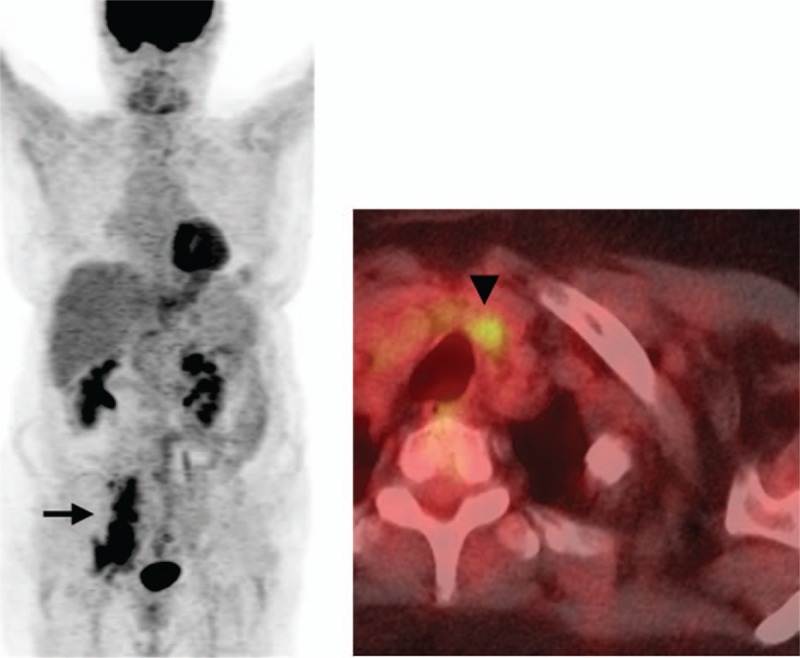
Figure 2.

A 71-year-old female patient with DLBCL undergoing a PET/CT for initial staging. The outside report indicated hypermetabolic right iliac chain adenopathy (black arrow on MIP image) and a cervical central compartment adenopathy (black arrowhead on axial fused PET/CT image) consistent with stage 3 disease. Second-opinion review at our institution reported infradiaphragmatic adenopathy consistent with stage 1 disease. The cervical hypermetabolic focus corresponds to a benign left thyroid nodule.
