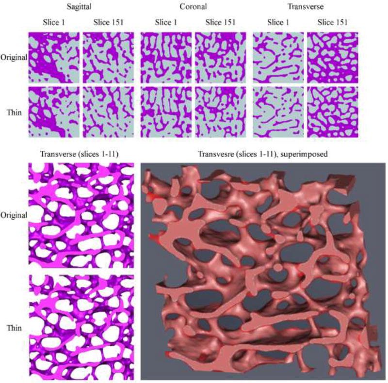
Figure 2.
The upper panel shows the first (#1) and last (#151) 2D slices of the original and thinned binarized cubical VOIs in the sagittal, coronal and transverse orientations. The left lower panel demonstrates a section of the 3D reconstructed cubes (original and thinned; slices #1–11) in the transverse direction. Comparing the two images reveals a significant decrease is trabecular thickness (and consequently an increase in trabecular spacing) throughout the sample. At the center bottom part of the images an originally intact trabecula (upper image) became severed (lower image). The right lower panel reveals the same section when the original and thinned reconstructions are superimposed. The light red areas are where both reconstructions represent ‘bone’. The semitransparent darker red represents areas which are ‘bone’ only in the original reconstruction (for a color version of this figure, please see the online version).

